


Oral Diagnosis
Good and accurate dental treatment can only be achieved by an accurate diagnosis. In dentistry, diagnosis is called “oral diagnosis” and can be described as “determination of all intraoral and extraoral problems while using scientific knowledge to find distinctions between them”.
Oral diagnosis bears importance not only with regard to dentistry but also in terms of systemic diseases; because some intra- and extra-oral findings can be indicators of certain systemic diseases.
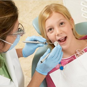
After the patient is seated in the dental chair, first, the doctor listens to the patient’s complaints. His/her account gives indications as to the characteristics of the problem. Gingival bleeding on brushing suggests a gingival disease; sensitivity to hot or cold suggests caries; mastication problems suggest missing teeth or toothache, or incompatible prostheses or a problem in the TMJ or mastication muscles.
A good oral diagnosis can be achieved by intraoral, extraoral, and radiological examinations.
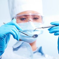
Extraoral examination is performed after listening the complaints of the patient in the dental chair and encompasses mouth and surrounding tissues (face, jaws, TMJ, lips, nose, neck, chin etc...). If any abnormality is detected in those tissues, further examination with CT (computed tomography) can be ordered.
A thorough intraoral examination should include inspection of gums, palate, tongue, and floor of mouth alongside teeth. Unusual findings in this area should be noted and if necessary, a comprehensive examination should be conducted. Gingiva should be examined in terms of both appearance (with bare eye) and volume (manually). If it is found to be redder or loose than normal, a gingival disease may be considered (gingivitis, periodontitis). If a swelling is detected during manual examination, abscess should come to mind. Findings obtained by bare eye examination, palpation, percussion etc. are assembled together and in combination with the complaints of the patient, placed in specially designed diagrams. For example, whether a complaint of pain is originated from caries or periodontal disease can be determined. Teeth are examined in a systematic order. They are evaluated in terms of gingival recession; plaque and calculus accumulation; mobility; caries; incompatible restorations; crowding; color, number, and shape problems. Following the examination of teeth one by one, dental occlusion pattern and relationship of jaws are checked.
Radiography verifies all the diagnostic examination noted above. Standard panoramic and left/right bite-wing radiographs provide information on unrecognized proximal caries, chronic lesions, impacted teeth, problems in the periodontal tissues (bone resorption), and formations in the jaw bones, overfilling, while revealing the status of permanent teeth, root formation, and resorption of deciduous teeth in children. Particularly in traumatized teeth, periapical radiographs taken at intervals act as a guide about the condition of tooth, root, and surrounding tissues. Moreover, taking a panoramic radiograph of every patient above 40 years age bears importance regarding detection of bone pathologies and clinically asymptomatic diseases.
Following detection of all the problems that are also supported by the radiographs, alternative treatment plans can be determined easily.
Shortly, half an hour spent during initial examination, leads to a perfect diagnosis followed by a successful and rational treatment.
Dentamar Ağız ve Diş Sağlığı Poliklinik Hizmetleri © 2025 | All Rights Reserved.